Latest News and Articles

Wellington Eye Centre’s Ophthalmologist
As we age certain eye conditions become more common, making it more important to have regular eye checks. After all, we rely heavily on our vision for independence and good quality of life. Not to worry – if diagnosed early, loss of vision can be minimised. We’ve put together some information on the most common age related eye conditions. Keep reading to learn more! Or, if you’d like to learn more about other common conditions check out this article on some of the most common eye conditions in New Zealand.
Cataracts
A cataract is a term used to describe the opacity or clouding of the natural lens of the eye. Cataracts are one of the common eye conditions, with cataract surgery being the most performed eye operation in New Zealand.
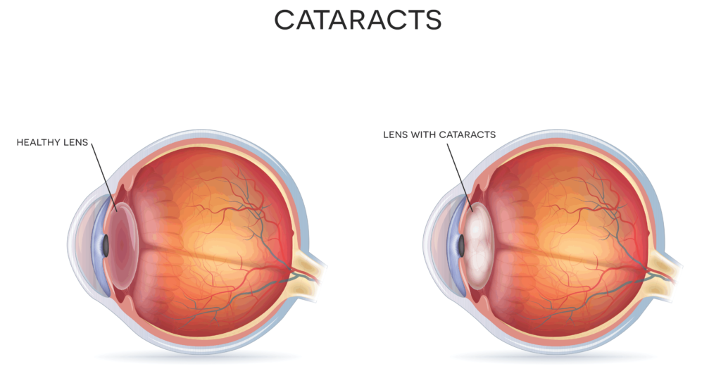
The lens is normally crystal clear in younger people but with increasing age, changes occur in the protein of the eye, causing it to develop a brownish discolouration. The lens is responsible for about 40% of the total focusing power of the eye. The remaining 60% of focusing is performed by the cornea, which is the front surface of the eye.
Initially this discolouration may have no effect on vision, but as it increases it can impair vision to the point where cataract surgery is required. Cataracts can affect vision by changing focus, causing blurring or clouding of vision, causing significant glare, halos and light sensitivity. By age 65, over 90% of people have signs of an early cataract and half of the people between the ages of 75 and 85 will have lost some vision due to a cataract.
What causes cataracts?
While a cataract is usually caused by ageing, cataracts can be congenital (when a child is born with cataracts). This is often a genetic or inherited condition, but can also be caused by non-genetic conditions such as congenital rubella syndrome. In some cases, cataracts can develop as a side effect of certain medications, particularly oral steroids such as prednisone. They may develop following eye injuries such as from a ball sport or a similar blow to the eye. Finally, cataracts may develop as a result of inflammation inside the eye (known as iritis or uveitis), or following surgery inside the eye such as glaucoma surgery and retinal detachment surgery.
Treatment for Cataracts
Cataract surgery is usually carried out when someone’s vision is compromised. There is a common misconception that cataract surgery cannot be performed until the cataract is “ripe” or severe. This dates back to a time when cataract surgery was a rather primitive operation and visual results were less than optimal. This meant that cataract surgery was often deferred as long as possible. Nowadays, cataract surgery is a very sophisticated, safe and effective operation. Cataract removal can be done at the point when vision is starting to be affected, rather than waiting until vision is severely impaired.
In most cases the surgery takes about 20 minutes and is carried out under local anaesthetic as a day case procedure. Normally vision is excellent the day after surgery. The risk of significant complications is very low. Cataract surgery is one of the safest eye operations carried out today.
Glaucoma
Glaucoma is the term used to describe a condition in which damage occurs to the optic nerve of the eye, usually as a result of high pressure inside the eye. The optic nerve is made up of retinal nerve fibres which travel from the retina, back to the visual processing areas in the brain. The optic nerve head is the structure at the back of the eye where the nerve fibres gather together along with blood vessels and connective tissue. These fibres then travel back to the brain via the optic nerve.
What causes Glaucoma?
Increasing pressure inside the eye damages the blood supply and nutrition in the optic nerve head. Less commonly, glaucoma can occur in people who have normal eye pressure. This is called normal-tension Glaucoma, and is not yet fully understood. Glaucoma may also occur following eye injuries, eye inflammation, from the use of steroid medications or eye drops, or in children as an inherited condition.
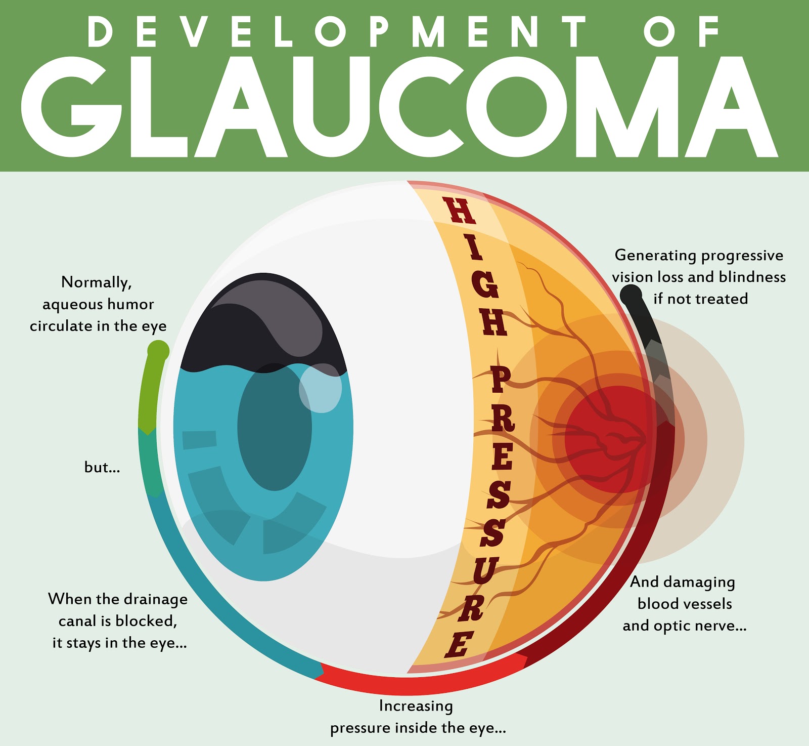
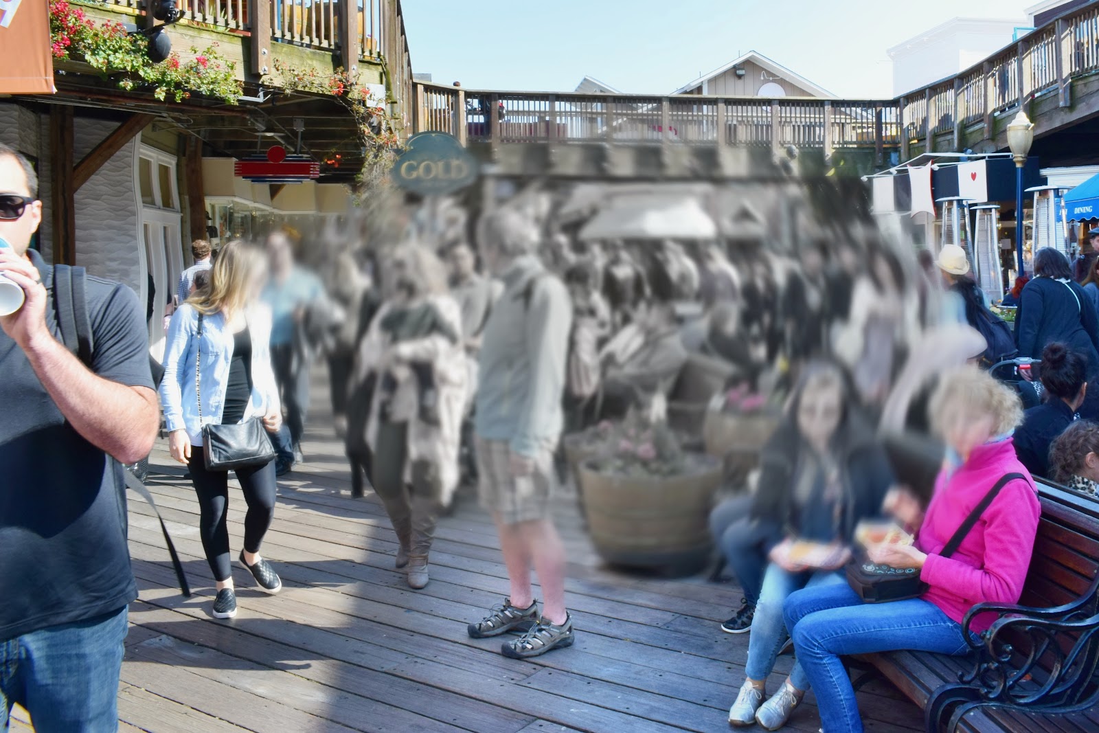
The damage caused by glaucoma at the optic nerve head can result in a progressive loss of peripheral vision, due to the damage of the retinal nerve fibres. In its early stages, glaucoma may only affect vision in the far periphery. This means somebody affected by glaucoma may be totally unaware of peripheral vision loss. Glaucoma is almost always a progressive condition. If it is left untreated it can cause progressive loss of vision to the point where only a small central island of vision remains (known as tunnel vision). So, it is important to have regular eye checks with an ophthalmologist or optometrist to diagnose early glaucoma.
There are two main types of glaucoma – chronic open angle glaucoma and acute angle closure glaucoma:
Chronic open angle glaucoma
This usually does not cause any symptoms in its early stages, this is due to it being a gradual increase in pressure and is not caused by narrowing of the drainage angle. The “open angle” refers to the drainage angle formed between the outer edge of the iris and the sclera. As glaucoma is usually progressive it can cause complete loss of vision if untreated, so it is important to have regular eye checks. High eye pressure and glaucoma becomes more common with increasing age, particularly over the age of 40 years.
Acute angle closure glaucoma
Acute angle closure glaucoma is also caused by high pressure inside the eye but is a completely different condition from chronic open angle glaucoma. In acute angle closure eye pressure can increase very rapidly to a high level. The condition causes severe pain in and around the eye and may also cause severe nausea and vomiting. If you have this type of glaucoma, you will certainly know about it! It’s an eye emergency because it can cause severe and permanent damage to vision in a very short time. The condition can be treated but this will need to be carried out by an ophthalmologist, usually in a hospital. Some people have a predisposition to developing acute angle closure glaucoma and this predisposition can be detected as part of a regular eye examination.

If you have a family history of glaucoma you should have your eyes checked more regularly, starting this at an earlier age than someone who does not have a family history. We would recommend getting your eyes checked every year or two if you have any of the risk factors.
Treatment for Glaucoma
Glaucoma treatment is designed to lower the eye pressure to the point where no further damage occurs to the optic nerve head. It’s very successful nowadays and the progression of the condition can be prevented in almost every case. Most patients who have glaucoma diagnosed will initially be treated with eye drops. These eye drops will need to be continued for life in most cases. They are a bit like medications for high blood pressure or high cholesterol – they are a treatment, not a cure.
A laser treatment for glaucoma called selective laser trabeculoplasty or SLT is frequently used. This may be used as a first-line treatment to avoid the need for eye drops or it may be used to give better control of the eye pressure in somebody who is already using eye drops. This is very different to the lasers used to correct vision.
Finally, if glaucoma continues to progress despite the use of eye drops and/or SLT, surgery may be used to treat the condition.
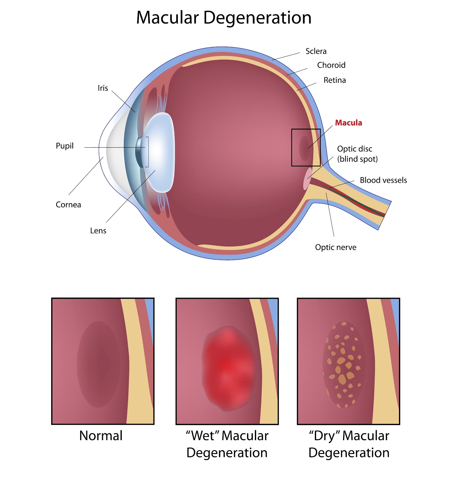
Age related macular degeneration
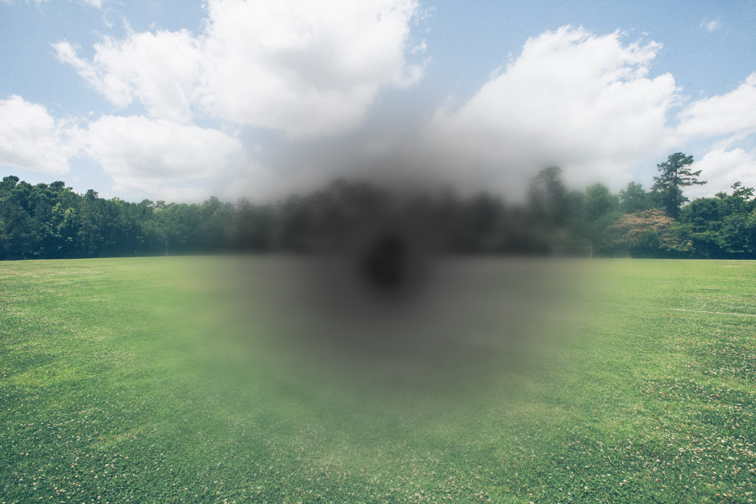
Macular degeneration or ARMD is a common eye condition in older people, which can cause significant damage to vision. It affects the macula, in the central part of the retina, caused by degeneration of macular cells. This results in a progressive loss of central vision.
Symptoms of Age-Related Macular Degeneration
Signs of the development of ARMD can be detected as part of a normal eye examination. Somebody who is developing ARMD may also notice changes in their central vision. These changes can include small blind spots in the central vision, distortion of straight lines, or objects look smaller or larger than expected. If you notice these changes you should have your eyes checked promptly by an ophthalmologist or optometrist.
Risk factors for the development of ARMD include increasing age, a family history of ARMD and smoking. In its early stages, ARMD may have little or no effect on vision. If vision is affected, this may be confused with reduced vision due to cataracts or as “just getting old”.
If you have signs of early ARMD you should ask your eye care provider about specific vitamin supplements that have been shown in large clinical trials to slow or reduce the progression of ARMD. There are also several treatments for ARMD which is affecting central vision. These include laser treatment and injections of medications known as “VEGF inhibitors”.
Presbyopia
Presbyopia is not really a “condition” at all – it happens to everyone when they get to middle age. One of the prices paid for living a normal life span. It’s the term used to describe the deterioration in close vision which occurs in all people once they reach middle age. The word presbyopia comes from the classical Greek words “Presbus” or old man, and “ops” or eye.
What causes Presbyopia?
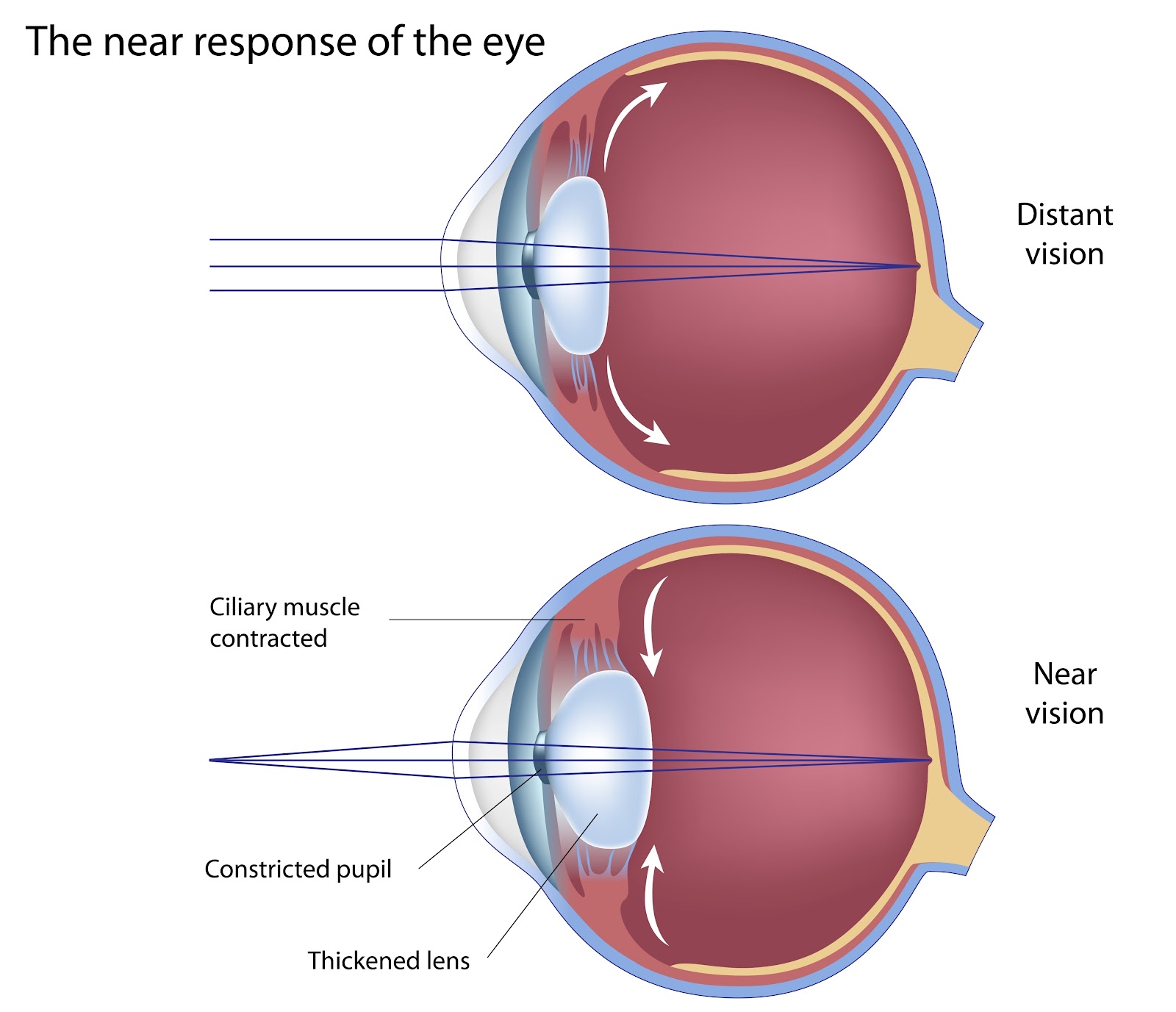
Presbyopia is caused by normal ageing changes that occur in the lens of the eye. The lens provides about 40% of the total focusing power of the eye and more importantly, it allows the eye to adjust focusing to allow clear vision at all distances.
For seeing at long distances, the lens adopts a thin shape with reduced curvature. Whereas for close distance vision such as reading, the lens becomes thicker with more curvature. In young people the lens is soft and flexible and can change rapidly to allow an easy change in focus at different distances. However, with increasing age, the lens becomes progressively stiffer and less flexible due to changes in the protein material that makes up the bulk of the lens. This process of stiffening starts to affect vision in the late 30s and becomes progressively worse up to the age of 60, at which time the normal human eye has lost all ability to focus at close distances.
Symptoms of Presbyopia
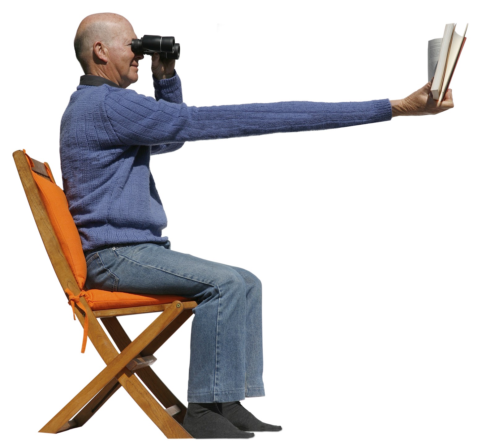
For many people, the first sign of the onset of presbyopia is difficulty changing focus from close to far. They may notice that distance vision is quite blurry after spending time reading or using a computer. As the condition progresses, the point of close focus stretches out and reading material will need to be held further away in order to see clearly. At some point reading glasses become necessary to bring the point of focus within arm’s length. Normally reading glasses must be strengthened every couple of years up to the age of 60.
People who are short sighted, that is who have vision that is clearer at close distances, can usually continue to read by taking their glasses off or not wearing their contact lenses. However, even these people usually eventually end up needing help to read; using what are known as progressive lenses in their glasses or reading glasses over their contact lenses.
Treatment for Presbyopia
Some fortunate people who have good distance vision in one eye and good near vision in the other may avoid needing reading glasses at all. This is the basis of a laser eye surgery procedure carried out at the Wellington Eye Centre called Presbyond Blended Vision. This procedure uses LASIK surgery to give one eye good vision in long distances and the other eye good vision at close distances.
This procedure works very well, and most people can expect to be completely or almost completely free of glasses or contact lenses. Of the patients who have had Presbyond Blended Vision surgery performed at the Wellington eye centre, about 96% of people do not wear glasses at all post-surgery. Approximately 4% of patients will still have a pair of glasses for occasional use at close distances or with small fiddly tasks such as threading a needle or reading very fine print. Some people are quite happy to correct their presbyopia with reading glasses or graduated lenses. For those people who can no longer be bothered with the hassle and expense involved with regular changes of glasses Presbyond Blended vision is an excellent alternative.
It’s important to get regular eye checks every one to two years when you are over the age of 40. If you are over the age of 65 or at a higher risk for any eye conditions then this increases to every six to 12 months.
Feel free to get in touch with the team at Wellington Eye Centre if you have any other questions or wish to book a consultation with Dr Logan. You can call us on 0800 733 327 or complete the contact form below.
Jul. 17, 2026 / Eye Health
What is Blepharitis?

May. 04, 2026 / Eye Health
Vitamins For Good Eye Health and Vision

Mar. 19, 2026 / Eye Health
Blue Light, Evil or Essential?
Feb. 10, 2026 / Eye Health
Age Related Macular Degeneration (ARMD)

Dec. 18, 2025 / Eye Health
Everything You Need To Know About Dry Eye
Jul. 17, 2026 | Eye Health
What is Blepharitis?

Jun. 16, 2026 | Laser Eye Surgery
PRESBYOND Recovery: What to Expect After Laser Blended Vision Surgery
May. 26, 2026 | Laser Eye Surgery
Why we recommend Dry Eye Treatments before your Laser Vision Correction
May. 04, 2026 | Eye Health
Vitamins For Good Eye Health and Vision

Apr. 16, 2026 | Laser Eye Surgery