Latest News and Articles
Wellington Eye Centre Ophthalmologist
The process of seeing is a hugely complex subject. To understand what happens when we “see” something, it is helpful to separate the subject into two parts. Firstly, the eye and how it functions, and secondly, how vision actually occurs, a process known as visual perception. Finally, we will show how damage to different parts of the visual system affects vision.
The reason for making this separation is that we do not “see” with just our eyes. We also see with our brains. If this sounds strange, think about dreaming. We see well in our dreams despite having our eyes closed and usually being in a darkened environment. People who experience lucid dreaming often describe seeing with far greater clarity and brilliance than in normal waking life. We need our eyes to see our waking environment, but they are not sufficient by themselves. We also need our brains to see.
A deeper look at the eye
Outer surfaces
The eye sits inside a bony cavity called the orbit or eye socket. The orbit contains the eye, the muscles that move the eye, blood vessels, nerves, and the tear glands. Fat surrounds these structures and acts as a shock absorber for the tissues.
The outer part of the eyeball consists of a tough white shell called the sclera (the white of the eye). This is made up of a strong connective tissue called collagen, which is also found in tendons and joints throughout the body. A thin transparent membrane called the conjunctiva covers the sclera. This runs to the edge of the cornea at the front of the eye and covers the back surface of the eyelids. Collagen makes up the cornea too, but unlike the sclera, it is normally crystal clear.
Inside the eye
Behind the cornea lies the iris, the coloured part of the eye. In the centre of the iris is a circular opening called the pupil. Contraction and relaxation of muscles in the iris cause the pupil to constrict or dilate in response to varying light levels. The space between the back of the cornea and the front of the iris is called the anterior chamber, filled with a clear salty fluid called the aqueous humor.
Immediately behind the iris sits the lens of the eye. The lens of the eye is responsible for adjusting the focusing of vision at different distances. Extremely fine fibres called zonules attach the lens to the ciliary muscle and hold it in place. When this muscle contracts or relaxes, tension on the zonules varies, which in turn allows the shape of the lens to change and alter the focusing of the eye.
A specialised nerve tissue called the retina lines the back of the eye. The retina functions like the film on a camera or a sensor in a digital camera; it detects light and starts the process of seeing. Before light reaches the retina it must travel through the vitreous humor which is a clear gel-like fluid. Which makes up a large portion of the eye. Occasionally a substance will enter the vitreous humour and remain suspended. You may see these as squiggly lines or black dots. We call these floaters.

How do we process the image?
The eye is often said to work like a camera. Though this is not entirely true for a film camera. Which use film to collect an image, which then must be developed outside the camera. However, digital cameras provide a good way of understanding how the eye works.
The lens focuses light and adjusts the focus so that objects at different distances are clear. The amount of light getting into the camera is adjusted using a diaphragm which can be opened or closed. This adjusts the amount of light reaching the image sensor at the back of the camera. The sensitivity of the sensor can also be adjusted so that it is more or less sensitive to light.
The image sensor has millions of individual pixels which are sensitive to different colours – red, green, or blue. These pixels produce a digital signal when hit by light photons. The firmware in the camera or software on a computer processes the final image.
How do our eyes focus?
The eye focuses in a similar way to a digital camera. In the eye, the cornea does about 60% of the total focusing, the lens then carries out the remaining 40%. The lens changes shape to focus the eye, becoming thicker to focus at close distances and thinner for far. The iris has a hole in its centre called the pupil. The pupil varies in size to adjust the amount of light entering the eye; narrowing in bright light, and enlarging in low light. This provides a rapid response to varying light levels.
After passing through the cornea, pupil and lens; light is then focused on the retina. This is the place where the process of “seeing” really begins. An enormous number of specialised nerve cells make up the retina and it is an extension of the brain. These cells detect incoming light, adjust to varying light levels, and perform some initial processing of visual information. The light-detecting cells are called photoreceptors.
There are 2 basic types of photoreceptor cells; rods and cones. Rod cells are in the peripheral part of the retina, they are responsible for night vision and peripheral vision. Rod vision is “black and white” and low resolution; a bit like a cheap camera. In a process called “dark adaptation”, rods can progressively increase their sensitivity to light in dark conditions. Dark adaptation is a relatively slow process, taking several minutes. Whereas cones are in the centre part of the retina, they are responsible for detailed, colour vision. Like a digital camera, there are 3 types of cones to detect different colours – red, green, and blue. Cones do not work well in poor lighting, which is why we progressively lose colour vision as it gets darker.
How does the image travel to our brain?
Photoreceptors contain a pigment that is sensitive to light. When light hits a photoreceptor, the pigment changes shape and this, in turn, causes an electrical signal to be produced. Photoreceptors connect to other nerve cells within the retina, these eventually connect to nerve fibres on the inner surface of the retina. The nerve fibres join together to form the optic nerve at the back of the eye.
The optic nerve leaves the eye socket and enters the brain cavity. The two nerves from each eye meet and join in a structure called the optic chiasm. The optic nerve from each eye then divides in the optic chiasm. Half of the nerve fibres from each side cross over and continue to the back of the brain, to an area of the brain called the visual cortex. As a result, the right side of the brain receives information from both optic nerves for the left visual field, and the left side of the brain receives information from both optic nerves for the right visual field.
How does our brain process this image?
Visual perception (the process of becoming aware of seeing something) occurs in the visual cortex. However significant processing of visual information occurs before signals from the eye reach the visual cortex. Most nerve fibres from the retina travel via the optic nerve to the lateral geniculate nucleus (LGN), an area inside the thalamus. The LGN is the main connection between the optic nerve and the occipital lobe. Here information from the retina separates into two streams, one containing information about colour and fine detail and the other containing contrast and motion information. The optic radiation pathways carry these two streams of information to the visual cortex. This is where integration of the information occurs, and the experience of vision is generated.
The optic nerve also carries the information needed for the mechanics of vision to two other sites in the brain. One site is a collection of cells that control the size of the pupil in response to varying light levels. Another site processes information concerning moving targets and information governing scanning of the eyes.

How does damage to the visual pathway affect vision?
The visual system can be damaged in many ways; including a congenital condition, injury, tumour or surgery. How vision is affected is determined by the injury location.
Loss of an eye or damage to the optic nerve
When one eye or optic nerve is lost, effects include loss of stereoscopic (3-D) vision and some loss of peripheral vision on the side of the lost eye. Typically, about 20% of peripheral vision will be lost on that side. This can cause difficulties moving in crowded environments and being aware of hazards coming from the blindside.
Loss of 3D vision can cause more significant problems. To have 3D vision, you need two functioning eyes. Sudden loss of vision in one eye can cause severe difficulties with depth perception, especially at close distances. Tasks such as walking down stairs and pouring drinks into a glass can be challenging. With time adaptation occurs; the brain is able to use what are known as “monocular clues” to judge depth. This adaptation occurs more quickly at a younger age, children who are blind in one eye may show no signs of having reduced depth perception. Practically speaking, one-eyed people are not limited by their lack of 3D vision.
Damage to the optic chiasm
Each eye and optic nerve is completely separate from the other side and damage to an eye or optic nerve will normally only affect vision on that side. However, the visual pathways from each eye become mixed together at the optic chiasm. From the optic chiasm back to the visual cortex damage to one side will affect vision from both eyes. At the chiasm, each optic nerve effectively divides in half. The side of the nerve that carries visual information from the nasal, or inside, half of the visual field stays on the same side of the brain. The other half of the nerve which carries information from the outside, or temporal, half of the visual field crosses over at the chiasm. This means that the left side of the visual pathway carries information from the right and the left eye, as does the right side.
The optic chiasm sits just in front of a small organ called the pituitary gland. If the pituitary gland develops a tumour or swelling, it can press on the optic chiasm. This pressure can damage nerve fibres which crossover in the chiasm, the ones that carry signals from the temporal or outside visual field from each eye. Temporal vision in both eyes can be lost causing a condition known as bitemporal hemianopia. This is often the first and only sign of a developing growth in the pituitary gland.
People with albinism have an abnormality of the chiasm where almost all of the nerve fibres cross over, resulting in defective mixing of information from each eye. This causes poor 3D vision.
Damage between the chiasm and the visual cortex
After the chiasm, information from both eyes is mixed together. The right visual pathway carries information from the left half of the visual field for each eye and vice versa for the left pathway. Therefore, damage to the right visual pathway will cause loss of vision on the left side. This is known as homonymous hemianopia; common causes of this can include strokes and brain tumours. Because there are so many other important structures in the same area as the visual pathway, other brain functions such as controlling speech and movement are likely to be affected.
Damage to the visual cortex
Damage to the visual cortex can affect one or both sides of the brain. The right and left visual cortexes sit right next to each other, so severe damage can affect both sides. Causes of damage to the visual cortex include tumours, head injury or a stroke.
If the visual cortex on one side of the brain is damaged, vision will be lost from the opposite half of the visual field from each eye. So if the left visual cortex is damaged there will be a loss of vision on the right side of each eye, again causing homonymous hemianopia. If the visual cortices on both sides are damaged or destroyed, all vision can be lost. This is known as cortical visual impairment.
Blind Sight
Extensive research has been carried out, investigating vision in people who suffer from cortical visual impairment. It has been found that these people show something called “blindsight”. Even though they are effectively totally blind and unable to “see” or perceive anything at all, they can quite accurately detect shapes and movements when test objects are placed in their “blind” visual fields. Despite being able to do this, they are completely unaware of seeing anything at all. The reason that this occurs is thought to be due to visual processing that has occurred in parts of the visual pathway, such as the LGN which are able to detect shape and movement. This processing occurs below the level of conscious awareness, however, and the person is not aware of having seen anything.
As you can see the eye is an incredibly complex organ. There are many different components to how we see in the brain too. Hopefully you’ve learned a thing or two along the way.
At the Wellington Eye Centre we love writing about eyes. We have some more articles you might like here:
- Viruses and the eye
- 10 Interesting facts about eyes
- Vitamins for good eye health and vision
- The truth behind common laser eye surgery myths
- Spring has almost sprung, how to look after your spring-related eye issues
Feel free to get in touch with the team at Wellington Eye Centre if you have any other questions or wish to book a consultation with Dr Logan. You can call us on 0800 733 327 or complete the contact form below.
May. 14, 2025 / Eye Health
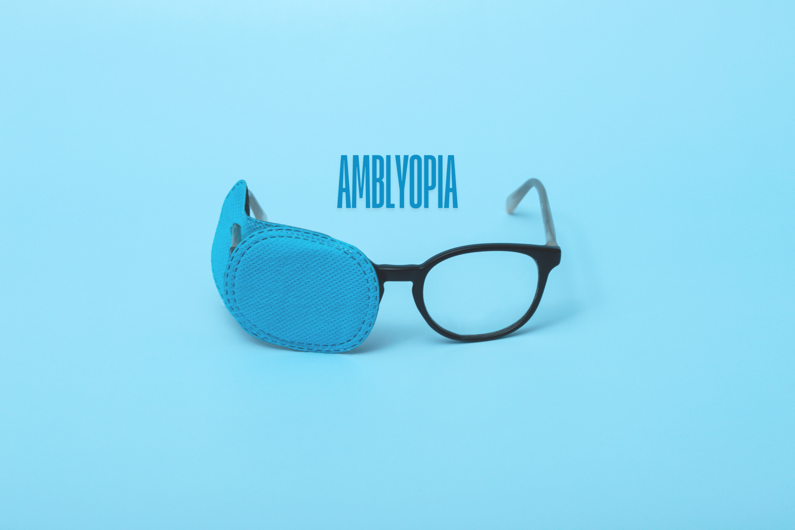
Amblyopia and Laser Eye Surgery

Dec. 15, 2024 / Eyesight
What are the visual requirements to join the Police in NZ?
Apr. 30, 2024 / Eyesight
What Can Go Wrong During Your Laser Eye Surgery?

Oct. 04, 2023 / Eyesight
What are the visual requirements for flying, set by the Civil Aviation Authority?
Sep. 20, 2023 / Eyesight
What happens when your vision after laser vision correction surgery is not what you expected?

Jun. 16, 2026 | Laser Eye Surgery
PRESBYOND Recovery: What to Expect After Laser Blended Vision Surgery

May. 26, 2026 | Laser Eye Surgery
Why we recommend Dry Eye Treatments before your Laser Vision Correction

May. 04, 2026 | Eye Health
Vitamins For Good Eye Health and Vision

Apr. 16, 2026 | Laser Eye Surgery
How Much Does Laser Eye Surgery Cost in 2026?

Mar. 19, 2026 | Eye Health